
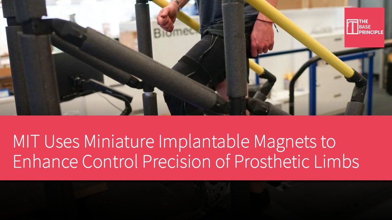
- MIT researchers demonstrate a miniature implanted magnet technology that enhances prosthetic control precision.
- The magnetic measurement method provides more accurate muscle movement information than traditional electromyography.
- The technology has been tested on three below-the-knee amputees, showing promising results.
- It could become a less invasive alternative for prosthetic control in the future.
Researchers at the Massachusetts Institute of Technology (MIT) have demonstrated a method using miniature implanted magnets to track muscle movements, which could help amputees control prosthetic limbs in a more natural manner. The technology has been tested on three below-the-knee amputees and shows promise in overcoming the limitations of current muscle signal systems. This method, known as magnetomicrometry, measures changes in the distance between magnetic beads implanted in the muscle. The researchers state that the muscle movement information provided by these measurements is more accurate and stable than traditional surface electrodes.
The related research findings have been published in Nature Sensors and build upon years of developing interfaces for prosthetic devices that allow them to respond more like natural limbs.
MIT's system is capable of tracking muscle movements to guide prosthetic control. Most current muscle-controlled prosthetic systems use electromyography (EMG), which measures electrical activity associated with muscle contractions. Electrodes placed on the skin detect these signals, which are then converted into commands for the mechanical limb by software. However, electrical activity does not directly reveal the mechanical behavior of muscles, including their length, speed of movement, and force-related characteristics. The reliability of these signals can also diminish during sustained muscle contractions, when sweat affects the electrodes, or when sensors pick up activity from nearby muscles.
Overcoming the Limitations of Traditional Electromyography
MIT's magnet-based approach aims to overcome these limitations by directly measuring muscle movements. Researchers implant a pair of miniature magnets into the muscle and place external sensors over the skin. As the muscle contracts or stretches, the distance between the magnets changes. The system tracks these changes to calculate the muscle's length and speed, allowing the prosthetic controller to guide limb movements.
These magnets, approximately 3 millimeters in diameter, are made from an alloy of neodymium, iron, and boron, and are coated with metal and polymer (parylene C) to improve biocompatibility. The researchers note that these magnetic signals can penetrate tissue to a depth of up to 6 centimeters.
The team tested this technology on three below-the-knee amputees, all of whom had undergone an antagonistic muscle neural interface (AMI) surgery. This surgical method reconnects opposing muscle groups, allowing them to continue communicating dynamically within the residual limb. This connection helps preserve proprioception, the ability to sense the position and movement of limbs. Maintaining this feedback can assist users in controlling their prosthetics more accurately. Participants received magnetic implants in up to four muscles associated with ankle movement. The researchers then compared magnetomicrometry with two established measurement methods: muscle-implanted electrodes and surface electrodes on the skin.
In the experiment, participants used muscle movements to control a cursor on a screen. The researchers converted the signals from each measurement system into control commands to compare the accuracy and consistency of different interfaces in supporting the task. However, the experiment did not involve participants walking with prosthetics controlled by the magnetic method; instead, the cursor task served as a test of the underlying control interface.
Potential Applications and Future Research
This research builds on earlier work by the MIT team, which demonstrated magnetomicrometry in animal models in 2021. The researchers have also developed prosthetic systems using implanted electrodes, including a mechanical knee designed to improve control through more direct measurement of muscle activity. This magnetic method could provide a less invasive alternative to implanted electrodes. Potential applications include mechanical ankle-foot prosthetics that can respond more naturally to the wearer’s intended movements. The researchers are currently conducting larger-scale studies to evaluate the effectiveness of using magnetic measurement to control prosthetics.
Further testing will be needed to determine the safety, durability, and performance of the system in daily activities. If these studies prove successful, magnetomicrometry could help bridge the gap between the movements people intend to make and the actions their prosthetics can reliably perform.
The Impact of Magnetomicrometry on Prosthetic Control
MIT's miniature implantable magnet technology showcases a new breakthrough in prosthetic control. Traditional electromyography methods suffer from low signal reliability, while magnetomicrometry offers more accurate data by directly measuring muscle movements, which has significant implications for amputees' daily lives. Through this technology, prosthetics can respond more naturally to users' intentions, enhancing the user experience. With further research and testing, this technology has the potential to become a mainstream option for future prosthetic control and may lead to advancements in related medical technologies.